Project No. 2485
STANDARD PROJECT
Primary Supervisor
Dr Wei-Feng Xue – University of Kent
Co-Supervisor(s)
Prof Louise C Serpell – University of Sussex
Summary
Atomic force microscopy (AFM) is a powerful and increasingly accessible technology that has a wide range of imaging applications, including nano-scale imaging of biological macromolecules, membrane surfaces and supramolecular assemblies.
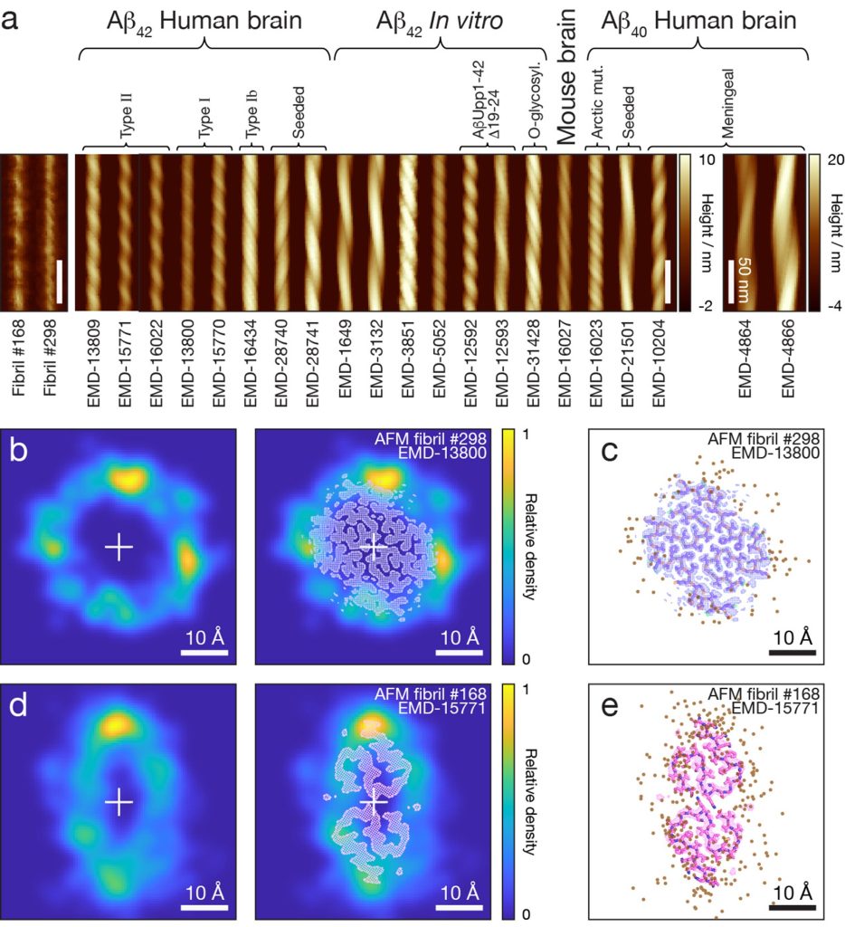
AFM is capable of producing detailed three-dimensional topographical height images with a high signal-to-noise ratio. This is a key capability of AFM, which enables the structural features of individual molecules to be studied without the need for molecular averaging, and could offer structural analysis applications where heterogeneity of molecular populations, structural variations between individual molecules, or population distribution properties in general, hold important information. However, the use of AFM imaging for general structural analyses of molecules such as biological macromolecules and assemblies has been very limited, especially in comparison to the recent advances of cryo-electron microscopy (cryo-EM) methods. Here, we will develop new methods for high-resolution structural analysis of individual molecules using AFM as a rapid and accessible structural analysis technology. Experimental AFM workflow and software image analysis and 3D structural reconstruction algorithms will be developed and tested by analysis of individual amyloid assemblies found in human dementia patients.
This project will, therefore, transform the use of AFM in structural biology, and in integrated methodologies together with other structural analysis tools to understand the structures and behaviours at individual molecule level. This project is particularly suited for candidates with biochemistry or biophysics background interested in developing transformative technologies focusing on molecular imaging and image analysis, structural biology methods or computational and data science approaches to biological challenges.

